Lower Extremities
This section of the documentation is under development
This section is being updated
Lower Extremity Components Identifier Overview
| Component Group | Identifier Range (Start) |
|---|---|
| Bones | 701000 |
| Knee joint structures | 702000 |
| Feet structures | 704000 |
| Leg soft tissues | 705000 |
| Tibiofibular structures | 706000 |
Bones
Femur
The femur cross section was optimised to meet the target values of Klein et al. (2015)1. The following target values were used (applying the regression model described in the paper and using age, stature and BMI of the 50F VIVA + models (50 years, 161.6 cm, 24 kg/m^2) An elliptic inner shape was assumed, which is in line with medical images. However, if a proper inner geometry becomes available, this should be updated. The maximum difference to the reference is 3.3%.
Femur cross sectional area
| Bone cross sectional area [mm^2] | L1 | L2 | L3 | L4 | L5 |
|---|---|---|---|---|---|
| Target from Klein et al. for 50F | 361 | 310 | 303 | 255 | 199 |
| Measured values in VIVA+ 50F | 372 | 306 | 300 | 252 | 193 |
Femur Solid Mesh Quality
| Criteria | limit | % of failed elements | limit | % of failed elements |
|---|---|---|---|---|
| Aspect Ratio | < 10 | 0 | 3 | 6.84 |
| Skewness | > 60^{\circ} | 0.04 | >45^{\circ} | 3.26 |
| Warping | < 15 | 0.09 | <10 | 0.67 |
| Jacobian | <0.3 | 0 | >0.7 | 1.56 |
| Internal Angle | >160^{\circ} | 0.05 | >140^{\circ} | 2.71 |
| <20^{\circ} | 0 | <30^{\circ} | 0.14 |
Femur Shell Mesh Quality
| Criteria | limit | % of failed elements | limit | % of failed elements |
|---|---|---|---|---|
| Aspect Ratio | < 10 | 0 | 3 | 1.38 |
| Skewness | > 60^{\circ} | 0 | >30^{\circ} | 10.6 |
| Warping | < 15 | 0 | <7 | 1.77 |
| Jacobian | <0.3 | 0 | >0.7 | 0.22 |
| Internal Angle | >160^{\circ} | 0 | >135^{\circ} | 2.76 |
| <20^{\circ} | 0 | <45^{\circ} | 2.46 |
Cortical bone properties are based on Mirzaali et al. (2016)2. Subjects with diagnosed osteoporosis were excluded. Trabecular bone properties are based on Ding et al. (1997)3
Tibia
Trabecular bone properties are based on Ding et al. (1997)3.
Tibia Mesh Quality
| Criteria | limit | ** % of failed elements** | limit | % of failed elements |
|---|---|---|---|---|
| Aspect Ratio | < 10 | 0 | 3 | 3.28 |
| Skewness | > 60^{\circ} | 0.53 | >45^{\circ} | 3.08 |
| Warping | < 15 | 0.31 | <10 | 0.79 |
| Jacobian | <0.3 | 0 | >0.7 | 1.12 |
| Internal Angle | >160^{\circ} | 0.24 | >140^{\circ} | 3.85 |
| <20^{\circ} | 0.07 | <30^{\circ} | 0.89 |
Fibula
Bone cross section properties are reported in Matsuura et al. (1999)4. Bone wall thickness ranges from 2 to 4 mm.
Fibula Mesh Quality
| Criteria | limit | ** % of failed elements** | limit | % of failed elements |
|---|---|---|---|---|
| Aspect Ratio | < 10 | 0 | 3 | 39.1 |
| Skewness | > 60^{\circ} | 0.47 | >45^{\circ} | 4.35 |
| Warping | < 15 | 0 | <10 | 0.11 |
| Jacobian | <0.3 | 0 | >0.7 | 0.70 |
| Internal Angle | >160^{\circ} | 0.13 | >140^{\circ} | 6.22 |
| <20^{\circ} | 0.04 | <30^{\circ} | 0.74 |
Patella
The patella is currently modelled as rigid.
Patella Solid Mesh Quality
| Criteria | limit | % of failed elements | limit | % of failed elements |
|---|---|---|---|---|
| Aspect Ratio | < 10 | 0 | 3 | 0 |
| Skewness | > 60^{\circ} | 0 | >45^{\circ} | 3.70 |
| Warping | < 15 | 0 | <10 | 3.70 |
| Jacobian | <0.3 | 0 | >0.7 | 29.6 |
| Internal Angle | >160^{\circ} | 0.62 | >140^{\circ} | 14.8 |
| <20^{\circ} | 0 | <30^{\circ} | 0 |
Patella Shell Mesh Quality
| Criteria | limit | % of failed elements | limit | % of failed elements |
|---|---|---|---|---|
| Aspect Ratio | < 10 | 0 | 3 | 0 |
| Skewness | > 60^{\circ} | 0 | >30^{\circ} | 7.22 |
| Warping | < 15 | 0 | <7 | 1.67 |
| Jacobian | <0.3 | 0 | >0.7 | 0 |
| Internal Angle | >160^{\circ} | 0 | >135^{\circ} | 4.44 |
| <20^{\circ} | 0 | <45^{\circ} | 3.33 |
Joints
Knee Joint Materials
Ligaments
The major knee ligaments are modeled as discrete beam elements.
Number of beams for each modeled knee ligament
| Ligament | Number of elements |
|---|---|
| MCL | 4 |
| LCL | 4 |
| aACL | 1 |
| pACL | 1 |
| aPCL | 1 |
| pACL | 1 |
| PL | 4 |
The knee ligament material properties are based on van Dommelen et al. (2005)5 and Kunitomi et al. (2017)6 and are modelled as discrete springs. Material properties for the patellar ligament are derived from Muller et al. (2004)7.
Ligament pretension
All knee ligaments are being pretensioned at the start of the simulation based on the value of pretension strain from Adouni et al. (2020)8 for the standing human.
The amout of ligament pretension is calculated as a difference between relaxed length of the ligaments (zero strain) and the distance between the ligament attachment nodes at the start of the simulation. The ligament pretensioning is performed in the first time step of the simulation (or in maximum of 1 ms) using a ramp up function.
Ligament pretension of Viva+ models
| Ligament | Initial strain [mm] |
|---|---|
| MCL | 3.3 % |
| LCL | 3.0 % |
| aACL | 4.9 % |
| pACL | 4.9 % |
| aPCL | 3.0 % |
| pACL | 3.0 % |
| PL | 1.8 % |
Knee Cartilage
Cartilage thickness is based on Faber et al. (2001)9 and Eckstein et al. (2001)10. The material properties are based on Robinson et al. (2016)11.
Meniscus
The material parameters are based on Peña et al. (2005) 12. An Ogden material model is applied using an alpha of 1 and therefore using Neo-Hookean modelling with a modulus of 59 MPa. The input parameters for meniscus were determined from the review by Joao in Trad et al. (2018)13.
Both cartilage and meniscus are modelled as linear elastic homogeneous materials, due to instant loading conditions as in Peña et al. (2006)14.
Connection between Fibula and Tibia
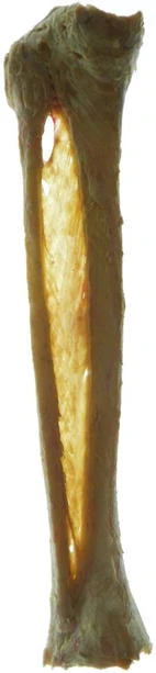
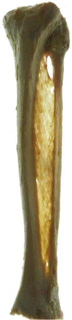
Crural Interosseous membrane (Anterior view, Posterior view)
140 beams were created oriented as described in Elamrani et al. (2013)15: "Fibers of the anterior layer made an angle of 13° (SD 2.6) with the axis of fibula. Those of the posterior layer made an angle of 24.2° (SD 2.48) with the axis of fibula." The average thickness of the membrane is 0.54 mm.
Stiffness of anterior tibiofibular ligament is based on Hoefnagels et al. (2007)16: 162 ± 64 N/mm.
Stiffness in fiber direction is assumed based on Minns and Hunter (1976)17. For the 2 mm x 20 mm sample an ultimate stress of 920 ± 205 Kgf/cm^2 = 0.09022118 GPa is reported at 7.7 %.
Assuming linear stiffness up to the ultimate stress, a young modulus of 1.17 GPa can be assumed for a sample with a cross section of 2 x 0.54 = ~1mm^2.
For the ligaments connecting the proximal end, a higher stiffness was assumed. It is currently set to 5 GPa.
Knee joint geometry
Attachment points on femur:
Blumensaat’s line (roof of femoral intercondylar): based on Iriuchishima et al. (2015)18. Ligaments were attached to the bones based on the anatomic landmarks described in the review of Bedi et al. (2018)19. Furthermore, the OpenKnee model was used as reference.
Ligament dimensions anteroposterior
| Ligament | Length [mm] |
Width [mm] |
Thickness [mm] |
CrossectionArea [mm^2] |
Sources |
|---|---|---|---|---|---|
| ACL | f:30.25, m:32.9 | f: 9.9, m: 12.2 | 4.78-4.89 | f:37.08, m:50.36 | 20^,21^,22'23'24 |
| ACL (tibial insertion) | - | f: 13.2, m: 13.5 (medio-lateral) | f:18.7, m: 20 (anteroposterior) | f: 118.85, m:142.5 | 22^,23 |
| ACL (femoral Insertion) | - | f: 6.3, m: 6.8 (anteroposterior) | f: 12.4, m: 14.4 (proximal-distal) | f: 81.45, m:98.9 | 22^,23 |
| PCL | 32-38 | 8-19.5 (mean=13.75) | 3.85-6.63 | 64.05 | 24^,25 |
| PCL (tibial insertion) | - | 9.58 | 9.12 | 147.67 | 24^,25^,26 |
| PCL (femoral insertion) | - | 5.35 | 20.69 | 148.2 | 24^,25^,26 |
| MCL | 87.5 | 10.9 (prox), 17.7 (mid), 10.7 (dist) | 2.1 | - | 27^,28^,29^,30 |
| MCL (femoral ins) | - | 11.5 (anteroposterior) | 9.2 (proximal-distal) | 75.5 | 27^,28^,31 |
| MCL (tibial ins sMCL) | - | 12.2 (anteroposterior) | 23.87 (proximal-distal) | 307.7 | 27^,28^,31 |
| MCL (tibial ins.- dist.) | - | 18 | 5 | 63.4 | 27 |
| LCL | f: 57.3, m:61.3 | 5.13 | 2.4 | - | 32^,33^,34 |
| LCL (femoral ins) | - | 9.7 (anteroposterior) | 11.2 | 52.1 | 32^,33^,35^,36 |
| LCL (tibial ins) | - | 7.97 (anteroposterior) | 11.9 | 38.6 | 32^,33^,35^,36 |
Lateral Collateral Ligament (LCL)
LaPrade et al. (2003)37: "The average cross-sectional area of the fibular collateral ligament attachment site on the femur was 0.48 cm^2 (range from 0.43 to 0.52).
Femoral attachment
According to Kamath et al. (2010)38, the femoral LCL insertion(black dot) is 58 ± 4.7 % across the width of the lateral femoral condyle along the Blumensaat line and 2.3 ± 2.3 mm distal to this point.
Fibular attachement
Based on LaPrade et al. (2003)37: "As the fibular collateral ligament coursed distally and attached on the lateral aspect of the fibular head, its average attachment was 8.2 mm (range, 6.8 to 9.7) posterior to the anterior margin of the fibular head and 28.4 mm (range, 25.1 to 30.6) distal to the tip of the fibular styloid process (Table 1). The average cross-sectional area of the attachment on the fibular head was 0.43 cm^2 (range, 0.39 to 0.50). The fibular collateral ligament attachment was, on average, 38 % (range, 28 % to 46 %) of the total width of the fibular head (anterior to posterior) from the anterior edge of the fibular head. The majority of the distal attachment was found in a bony depression that extended to approximately the distal one-third of the lateral aspect of the fibular head (Figs. 1 and 2). The remaining fibers extended further distally along with the peroneus longus fascia.25,26 The average total length of the fibular collateral ligament between its attachment sites was 69.6 mm (range, 62.6 to 73.5)."
Medial Collateral Ligament (MCL)
Based on Wijdicks et al. (2009)39 - values from table 5 will be used.
Additionally, figure from Prince et al. (2015)40 was used.
Femoral attachment
MCL attachment and sMCL attachment
"Line 1 is an extension of the posterior femoral cortex, and line 2 is drawn perpendicular to line 1, intersecting the most posterior aspect of the Blumensaat line" "The femoral attachment of the sMCL was found to be, on average, 8.6 mm anterior to the posterior femoral cortex line and 11.0 mm distal to the intersection of the posterior femoral cortex line (line 1) and the line intersecting the posterior aspect of the Blumensaat line (line 2)." 40
Tibial attachment
"On the lateral tibial radiographs, the proximal and distal tibial attachments of the superficial medial collateral ligament were 15.9 ± 5.2 mm and 66.1 ± 3.6 mm distal to the tibial inclination, respectively." 40
Anterior Cruciate Ligament (ACL)
Harner et al. (1999)41 (from Bedi et al. (2018)19) "The ACL is formed by 2 main bundles, the anteromedial (AM) and posterolateral (PL) bundles, which are named for their tibial insertions and provide the primary restraint against anterior tibial translation and secondary restraint against internal tibial rotation, respectively. It is 31±2 mm long with a mean diameter of 10±2 mm, although it fans out at the insertions to approximately 3.5 times the midsubstance width 7."
The distance between the attachment point in the baseline seated VIVA+ model is 31 mm.
Femoral insertion
ACL attachment point on femur is determined based on the radiographic quadrant mehtod: "distance t (representing the total sagittal diameter of the lateral condyle measured along Blumensaat's line), distance h (representing the maximum intercondylar notch height), distance a (representing the distance of point K from the most dorsal subchondral contour of the lateral femoral condyle), and distance b (representing the distance of point K from Blumensaat's line). Distance a is a partial distance of t and distance b is a partial distance of h, and distances a and b are expressed as length ratios of t and h. The center of the femoral insertion of the ACL was located at 24.8 % of the distance t measured from the most posterior contour of the lateral femoral condyle and at 28.5% of the height h measured from Blumensaat's line. Based on these results, the ACL can be found just inferior to the most superoposterior quadrant, which means in anatomic terms it is localized from the dorsal border of the condyle at approximately a quarter of the whole sagittal diameter of the condyle and from the roof of the notch at approximately a quarter of the notch height."
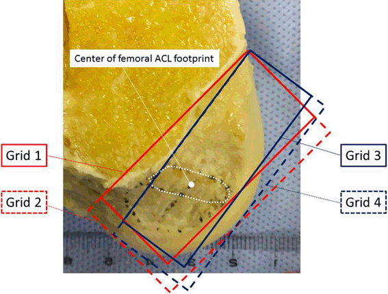
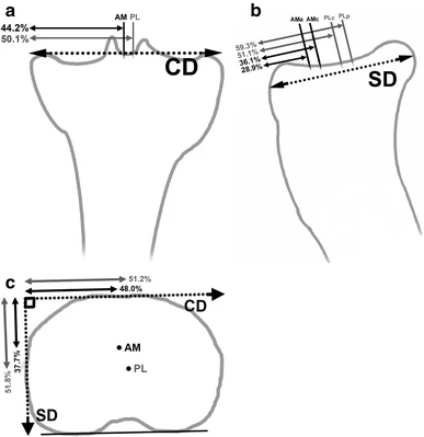
According to Yahagi et al. (2017)42, who are proposing a method which is applicable also for cases where the Blumensaat's line is not a straight line, the hill is excluded to derive the Blumensaat line (grid 1) (ACL footprint).
"In small hill type knees, the ACL center was placed as follows: Grid (1) 37.5 ± 6% in the shallow–deep, 50.2 ± 8.3% in the high–low directions. [..] In large hill type knees, the ACL center was placed as follows: Grid (1) 37.1 ± 5.6% in the shallow–deep, 50.4 ± 5.8% in the high–low directions"
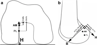
Method to derive both bundle attachments as in Pietrini et al. (2011)43:
Tibial insertion
Stӓubli and Rauschning (1994)44: 43.3% of the anterior-to-posterior distance across the tibia as measured at the level of the posterior tibial margin at the posterior intercondylar area.In their study,the anteriormost fibers inserted at 27.5% across the plateau.
Frank et al. (2010)45: "The average AP diameter of the tibia was measured to be 50 ± 4 mm (range 40–64 mm). Female knees averaged 47 ± 3 mm compared to 52 ± 4 mm in men. The anterior-most position of the ACL attachment on the tibia was, on average, 14 ± 3 mm (range 8–26 mm) from the anterior edge of the tibia, or 28 ± 5 % the total depth of the tibia. In women, the anterior-most position of the insertion was, on average, 13 ± 2 mm (28 ± 5 %) compared to 15 ± 3 mm (28 ± 5%) in men. The posterior-most position of the ACL attachment on the tibia was located, on average, 31 ± 4 mm (range 23–40) from the anterior edge of the tibia, or 63 ± 6% the depth of the tibia. In women, the posterior-most position was, on average, 29 ± 3 mm (62 ± 5%) contrasted to 33 ± 4 mm (64 ± 5 %) in men. Finally, the central portion of the ACL attachment on the tibia was located, on average, 23 ± 3 mm (range 16–30 mm). This center position corresponds to a point 46 ± 4 % of the total tibial AP diameter as described. In women, this position was located at 21 ± 2 mm (45 ± 4 %) compared to 24 ± 3 mm (46 ± 4 %) in men. It was determined that the ACL takes up an average 36 ± 6 % of the sagittal depth of the tibia and that the tibial insertion of the ACL is located between 28 and 63 % of the total depth of the tibia in the anterior–posterior (sagittal) plane."
Posterior Cruciate Ligament (PCL)
Positionining of the bundles is based on relative values provided in table 1, 2 and 3 of the Johannsen et al. (2012)46 publication.
The distance between the attachment point in the baseline seated VIVA+ model is 36 mm.
Meniscus
The average thickness of the medial meniscus is 2.55 mm according to Bloecker et al. (2011)47 (40 male and 62 female knees were measured in MRI) For males it should be 2.8 mm.
Data based on Bloecker et al. (2011)47: Table 1 and Table 2.
Meniscus width and thickness
| Mean avg. thickness [mm] |
Mean max. thickness [mm] |
Mean avg. width [mm] |
Mean max. width [mm] |
|
|---|---|---|---|---|
| Females Medial | 2.55 | 7.15 | 9.11 | 16.9 |
| Females Lateral | 2.51 | 6.75 | 8.60 | 18.8 |
| Males Medial | 2.80 | 7.71 | 9.93 | 12.5 |
| Males Lateral | 2.67 | 7.23 | 10.1 | 14.2 |
Knee Cartilage
Male vs. Female cartilage thickness, young healthy individuals, MRI based data on Faber et al. (2001)9
| Location | Female mean Thickness [mm] |
Male mean Thickness [mm] |
Female maximal Thickness [mm] |
Male maximal Thickness [mm] |
Female Area [mm^2] |
Male Area [mm^2] |
|---|---|---|---|---|---|---|
| Patella | 2.2±0.43 | 2.39±0.42 | 4.51±1.08 | 5.26±0.99 | 1047±123 | 1289±158 |
| Femur (total) | 1.79±0.22 | 1.88±0.29 | - | - | 5478±655 | 6554±391 |
| Trochlea | 2.01±0.25 | 2.05±0.32 | 4.2±0.48 | 4.51±0.72 | - | - |
| Medial condyle | 1.69±0.24 | 1.86±0.31 | 3.73±0.67 | 3.89±0.85 | - | - |
| Lateral condyle | 1.65±0.33 | 1.73±0.32 | 3.29±0.64 | 3.69±0.47 | - | - |
| Tibia medial | 1.2±0.19 | 1.36±0.15 | 2.9±0.92 | 3.43±0.86 | 811±122 | 1078±235 |
| Tibia lateral | 1.61±0.25 | 1.7±0.27 | 3.96±0.51 | 4.54±0.91 | 881±98 | 1175±147 |
| Knee total | 1.86±0.24 | 2.01±0.31 | - | - | 8218±795 | 10096±498 |
Cartilage: Gender specific dimensions by Eckstein et al. (2001)10
| Location | Female Thickness [mm] |
Male Thickness [mm] |
Female Area [mm^2] |
Male Area [mm^2] |
|---|---|---|---|---|
| Patella | 2.50 | 2.60 | 1100 | 1400 |
| Femur (total) | 1.60 | 1.75 | 5000 | 6500 |
| Tibia medial | 1.45 | 1.55 | 900 | 1150 |
| Tibia lateral | 1.75 | 2.00 | 900 | 1150 |
Patellar Ligament (PL)
The attachment point on the tibia is 36 mm below the most distal edge of the patella.
This is in line with Yoo et al. (2007)48, where an average value of 38 mm was reported from the 30 female knees which were measured in MRIs.
They report a proximal width of 27.5 mm and a tickness of 3 mm while for the distal tendon a tickness of 4.6 mm and a width of 21.5 mm is reported.
Patellar ligament dimensions
| Distal length [mm] |
Distal width [mm] |
Distal thickness [mm] |
Proximal width [mm] |
Proximal thickness [mm] |
|
|---|---|---|---|---|---|
| Female (Yoo et al. (2007)48) | 38 | 21.5 | 4.6 | 27.5 | 3 |
| VIVA+50F | 36 | 23.4 at patella; 25 at distal end | 4.6 | 26 at patella; 21 at most proximal | 3 |
| Male (Yoo et al. (2007)48) | |||||
| VIVA+50M |
Quadriceps muscle
For the muscles the material model »S15_MAT_SPRING_MUSCLE« has been used, which is defined for discrete beam elements with the possibility of activation. The model is described in LS-Dyna manual 49, by Hill et al. (1938) 50 and Winters et al. (1990) 51. Some parameters were also used from Audu et al. (1985)52, Horst et al. (2002)53, Mukherjee et al. (2007)54 and Arnold et al. (2009)55.
The main imput parametres are:
- initial length (L0) (depending on the individual model leg length)
- maximum shortening velocity (VMAX) 55^,53
- function of activation (𝑓A) (if used) 54
- peak isometric force (FMAX) 51^,55
- functions for : active tension vs. length function (𝑓𝑇𝐿) 49
- active tension vs. velocity function (𝑓𝑇V) 49^,52
- force vs. length function for parallel elastic element (𝑓PE) 49
- The the initial model configuration only one discrete element was used for the combination of four heads of quadriceps muscle. The input parameters for all four heads were summed up and used for the single discrete element.
Ankle joint
Simplified kinematic revolute joint for ankle is defined in the model between tibia-fibula and talus - rotational axis from lateral to medial malleolus - from Mansfield et al. (2018)56
Contact definitions
The contact definition for lower extremity joints is defined using a single "Automatic Single Surface" joint definition. It includes structures that can come in contact at the rotation of the joints (bone ends, cartilage, ligaments, capsules). In the internal knee joint contact the patella is excluded from the contact definition. For the contact between Femoral condyle, Tibial plateau, patella and outer skin an additional "Automatic Surface to Surface" contact was defined to get the correct kinematics of the patella and to prevent the protrusion of the patella through the skin.
Contact between bones and soft tissues
| Contact | Contact ID | Contact Type |
|---|---|---|
| Hip_Internal | 700000 | Automatic Single Surface |
| Knee_Internal | 700000 | Automatic Single Surface |
| Knee_External | 700010 | Automatic Surface To Surface |
| Ankle_Internal | 700000 | Automatic Single Surface |
Future model improvements
Average shapes of the bones based on statistical geometry.
Tuemer et al. (2018)57: Three-dimensional analysis of shape variations and symmetry of the fibula, tibia, calcaneus and talus.
Grant et al. (2020)58: Development and validation of statistical shape models of the primary functional bone segments of the foot.
References
-
Katelyn F. Klein, Jingwen Hu, Matthew P. Reed, Carrie N. Hoff, and Jonathan D. Rupp. Development and validation of statistical models of femur geometry for use with parametric finite element models. Annals of Biomedical Engineering, 43(10):2503–2514, mar 2015. doi:10.1007/s10439-015-1307-6. ↩
-
Mohammad J. Mirzaali, J. Jakob Schwiedrzik, Suwanwadee Thaiwichai, James P. Best, Johann Michler, Philippe K. Zysset, and Uwe Wolfram. Mechanical properties of cortical bone and their relationships with age, gender, composition and microindentation properties in the elderly. Bone, 93:196–211, dec 2016. doi:10.1016/j.bone.2015.11.018. ↩
-
M. Ding, M. Dalstra, C. C. Daniellsen, J. Kabel, I. Hvid, and F. Linde. Age variations in the properties of human tibial trabecular bone. J Bone Joint Surg Br., 1997. ↩↩
-
M Matsuura, K Ohno, K Michi, K Egawa, and R Takiguchi. Clinicoanatomic examination of the fibula: anatomic basis for dental implant placement. The International journal of oral & maxillofacial implants, 14:879–884, 1999. ↩
-
V. Characterization of the rate-dependent mechanical properties and failure of human knee ligaments. In SAE Technical Paper Series. SAE International, apr 2005. doi:10.4271/2005-01-0293. ↩
-
Shouhei Kunitomi, Yoshihiro Yamamoto, Ryosuke Kato, Jacobo Antona‐Makoshi, Atsuhiro Konosu, Yasuhiro Dokko, and Tsuyoshi Yasuki. The development of the lower extremity of a human fe model and the influence of anatomical detailed modelling in vehicle‐to‐pedestrian impacts. In International Research Council on the Biomechanics of Injury (IRCOBI). 2017. URL: http://www.ircobi.org/wordpress/downloads/irc17/pdf-files/62.pdf. ↩
-
Sérgio Swain Müller, Paulo Roberto de Almeida Silvares, Hamilton da Rosa Pereira, Marcos Augusto de Moraes Silva, Trajano Sardenberg, and Tomaz Puga Leivas. Análise comparativa das propriedades mecânicas do ligamento da patela e do tendão calcâneo. Acta Ortopédica Brasileira, 12(3):134–140, sep 2004. doi:10.1590/s1413-78522004000300001. ↩
-
Malek Adouni, Tanvir R. Faisal, and Yasin Y. Dhaher. Computational frame of ligament in situ strain in a full knee model. Computers in Biology and Medicine, 126:104012, nov 2020. doi:10.1016/j.compbiomed.2020.104012. ↩
-
S. C. Faber, F. Eckstein, S. Lukasz, R. Mühlbauer, J. Hohe, K.-H. Englmeier, and M. Reiser. Gender differences in knee joint cartilage thickness, volume and articular surface areas: assessment with quantitative three-dimensional MR imaging. Skeletal Radiology, 30(3):144–150, mar 2001. doi:10.1007/s002560000320. ↩↩
-
F. Eckstein, Maximilian Reiser, Karl-Hans Englmeier, and Reinhard Putz. In vivo morphometry and functional analysis of human articular cartilage with quantitative magnetic resonance imaging - from image to data, from data to theory. Anatomy and Embryology, 203(3):147–173, feb 2001. doi:10.1007/s004290000154. ↩↩
-
Dale L Robinson, Mariana E Kersh, Nicole C Walsh, David C Ackland, Richard N de Steiger, and Marcus G Pandy. Mechanical properties of normal and osteoarthritic human articular cartilage. Journal of the mechanical behavior of biomedical materials, 61:96–109, August 2016. doi:10.1016/j.jmbbm.2016.01.015. ↩
-
E Peña, B Calvo, M A Martínez, D Palanca, and M Doblaré. Finite element analysis of the effect of meniscal tears and meniscectomies on human knee biomechanics. Clinical biomechanics (Bristol, Avon), 20:498–507, June 2005. doi:10.1016/j.clinbiomech.2005.01.009. ↩
-
Zahra Trad, Abdelwahed Barkaoui, Moez Chafra, and João Manuel R.S. Tavares. FEM Analysis of the Human Knee Joint. Springer International Publishing, 2018. doi:10.1007/978-3-319-74158-1. ↩
-
E. Peña, B. Calvo, M.A. Mart\'ınez, and M. Doblaré. A three-dimensional finite element analysis of the combined behavior of ligaments and menisci in the healthy human knee joint. Journal of Biomechanics, 39(9):1686–1701, jan 2006. doi:10.1016/j.jbiomech.2005.04.030. ↩
-
Driss Elamrani, Aurélien Aumar, Guillaume Wavreille, and Christian Fontaine. Comparative morphometry of the antebrachial and crural interosseous membranes: preliminary study for the use of the crural interosseous membrane in the surgical repair of the antebrachial interosseous membrane tears. Surgical and Radiologic Anatomy, 36(4):333–339, sep 2013. doi:10.1007/s00276-013-1199-9. ↩
-
Eva M. Hoefnagels, Matthew D. Waites, Ian D. Wing, Stephen M. Belkoff, and Bart A. Swierstra. Biomechanical comparison of the interosseous tibiofibular ligament and the anterior tibiofibular ligament. Foot & Ankle International, 28(5):602–604, may 2007. doi:10.3113/fai.2007.0602. ↩
-
R. J. Minns and J. A. A. Hunter. The mechanical and structural characteristics of the tibio-fibular interosseous membrane. Acta Orthopaedica Scandinavica, 47(2):236–240, jan 1976. doi:10.3109/17453677608989725. ↩
-
Takanori Iriuchishima, Keinosuke Ryu, Shin Aizawa, and Freddie H. Fu. Blumensaat's line is not always straight: morphological variations of the lateral wall of the femoral intercondylar notch. Knee Surgery, Sports Traumatology, Arthroscopy, 24(9):2752–2757, mar 2015. doi:10.1007/s00167-015-3579-7. ↩
-
Asheesh Bedi, Robert F. LaPrade, and M. Tyrrell Burrus. Radiographic and anatomic landmarks of the major knee ligaments. The Journal of Bone and Joint Surgery, 100(14):1241–1250, jul 2018. doi:10.2106/jbjs.17.01135. ↩↩
-
Allen F. Anderson, David C. Dome, Shiva Gautam, Mark H. Awh, and Gregory W. Rennirt. Correlation of anthropometric measurements, strength, anterior cruciate ligament size, and intercondylar notch characteristics to sex differences in anterior cruciate ligament tear rates. The American Journal of Sports Medicine, 29(1):58–66, jan 2001. doi:10.1177/03635465010290011501. ↩
-
Naveen Chandrashekar, James Slauterbeck, and Javad Hashemi. Sex-based differences in the anthropometric characteristics of the anterior cruciate ligament and its relation to intercondylar notch geometry. The American Journal of Sports Medicine, 33(10):1492–1498, oct 2005. doi:10.1177/0363546504274149. ↩
-
Stephanie G. Cone, Danielle Howe, and Matthew B. Fisher. Size and shape of the human anterior cruciate ligament and the impact of sex and skeletal growth. JBJS Reviews, 7(6):e8–e8, jun 2019. doi:10.2106/jbjs.rvw.18.00145. ↩↩↩
-
Lazar Stijak, Vidosava Radonjić, Valentina Nikolić, Zoran Blagojević, Milan Aksić, and Branislav Filipović. Correlation between the morphometric parameters of the anterior cruciate ligament and the intercondylar width: gender and age differences. Knee Surgery, Sports Traumatology, Arthroscopy, 17(7):812–817, may 2009. doi:10.1007/s00167-009-0807-z. ↩↩↩
-
Eleni Triantafyllidi, Nikolaos K. Paschos, Anna Goussia, Nektaria-Marianthi Barkoula, Dimitrios A. Exarchos, Theodore E. Matikas, Vassiliki Malamou-Mitsi, and Anastasios D. Georgoulis. The shape and the thickness of the anterior cruciate ligament along its length in relation to the posterior cruciate ligament: a cadaveric study. Arthroscopy: The Journal of Arthroscopic & Related Surgery, 29(12):1963–1973, dec 2013. doi:10.1016/j.arthro.2013.09.007. ↩↩↩↩
-
Stephanie L. Logterman, Frank B. Wydra, and Rachel M. Frank. Posterior cruciate ligament: anatomy and biomechanics. Current Reviews in Musculoskeletal Medicine, 11(3):510–514, may 2018. doi:10.1007/s12178-018-9492-1. ↩↩↩
-
Masaaki Takahashi, Takamasa Matsubara, Mitsuhito Doi, Daisuke Suzuki, and Akira Nagano. Anatomical study of the femoral and tibial insertions of the anterolateral and posteromedial bundles of human posterior cruciate ligament. Knee Surgery, Sports Traumatology, Arthroscopy, 14(11):1055–1059, aug 2006. doi:10.1007/s00167-006-0192-9. ↩↩
-
Fang Liu, Bing Yue, Hemanth R Gadikota, Michal Kozanek, Wanjun Liu, Thomas J Gill, Harry E Rubash, and Guoan Li. Morphology of the medial collateral ligament of the knee. Journal of Orthopaedic Surgery and Research, 5(1):69, 2010. doi:10.1186/1749-799x-5-69. ↩↩↩↩
-
Norihiro OTAKE, Huayue CHEN, Xianfeng YAO, and Shizuko SHOUMURA. Morphologic study of the lateral and medial collateral ligaments of the human knee. Okajimas Folia Anatomica Japonica, 83(4):115–122, 2007. doi:10.2535/ofaj.83.115. ↩↩↩
-
Sang Eun Park, Louis E. DeFrate, Jeremy F. Suggs, Thomas J. Gill, Harry E. Rubash, and Guoan Li. Erratum to \textquotedblleft the change in length of the medial and lateral collateral ligaments during in vivo knee flexion\textquotedblright . The Knee, 13(1):77–82, jan 2006. doi:10.1016/j.knee.2004.12.012. ↩
-
William T. Wilson, Angela H. Deakin, Anthony P. Payne, Frederic Picard, and Scott C. Wearing. Comparative analysis of the structural properties of the collateral ligaments of the human knee. Journal of Orthopaedic & Sports Physical Therapy, 42(4):345–351, apr 2012. doi:10.2519/jospt.2012.3919. ↩
-
Mitchell I. Kennedy, Steven Claes, Fernando Augusto Freitas Fuso, Brady T. Williams, Mary T. Goldsmith, Travis Lee Turnbull, Coen A. Wijdicks, and Robert F. LaPrade. The anterolateral ligament. The American Journal of Sports Medicine, 43(7):1606–1615, apr 2015. doi:10.1177/0363546515578253. ↩↩
-
Espregueira-Mendes and M. Vieira da Silva. Anatomy of the lateral collateral ligament: a cadaver and histological study. Knee Surgery, Sports Traumatology, Arthroscopy, 14(3):221–228, oct 2005. doi:10.1007/s00167-005-0681-2. ↩↩↩
-
Brad R. Meister, Stanley P. Michael, Ray A. Moyer, John D. Kelly, and Carson D. Schneck. Anatomy and kinematics of the lateral collateral ligament of the knee. The American Journal of Sports Medicine, 28(6):869–878, nov 2000. doi:10.1177/03635465000280061601. ↩↩↩
-
Sebastian Tschauner, Erich Sorantin, Georg Singer, Robert Eberl, Annelie-Martina Weinberg, Peter Schmidt, and Tanja Kraus. The origin points of the knee collateral ligaments: an MRI study on paediatric patients during growth. Knee Surgery, Sports Traumatology, Arthroscopy, 24(1):18–25, apr 2016. doi:10.1007/s00167-014-2991-8. ↩
-
J.-M. Brinkman, P. J. A. Schwering, L. Blankevoort, J. G. Koolos, J. Luites, and A. B. Wymenga. The insertion geometry of the posterolateral corner of the knee. The Journal of Bone and Joint Surgery. British volume, 87-B(10):1364–1368, oct 2005. doi:10.1302/0301-620x.87b10.16536. ↩↩
-
Young-Bin Song, Koichi Watanabe, Elizabeth Hogan, Anthony V. D\textquotesingle Antoni, Anthony C. Dilandro, Nihal Apaydin, Marios Loukas, Mohammadali M. Shoja, and R. Shane Tubbs. The fibular collateral ligament of the knee. Clinical Anatomy, 27(5):789–797, nov 2013. doi:10.1002/ca.22301. ↩↩
-
Robert F. LaPrade, Thuan V. Ly, Fred A. Wentorf, and Lars Engebretsen. The posterolateral attachments of the knee. The American Journal of Sports Medicine, 31(6):854–860, nov 2003. doi:10.1177/03635465030310062101. ↩↩
-
Ganesh V. Kamath, John C. Redfern, and Robert T. Burks. Femoral radiographic landmarks for lateral collateral ligament reconstruction and repair. The American Journal of Sports Medicine, 38(3):570–574, mar 2010. doi:10.1177/0363546509350066. ↩
-
Coen A. Wijdicks, Chad J. Griffith, Robert F. LaPrade, Stanislav I. Spiridonov, Steinar Johansen, Bryan M. Armitage, and Lars Engebretsen. Medial knee injury. The American Journal of Sports Medicine, 37(9):1771–1776, jul 2009. doi:10.1177/0363546509335191. ↩
-
Matthew R. Prince, Andrew J. Blackman, Alexander H. King, Michael J. Stuart, and Bruce A. Levy. Open anatomic reconstruction of the medial collateral ligament and posteromedial corner. Arthroscopy Techniques, 4(6):e885–e890, dec 2015. doi:10.1016/j.eats.2015.08.013. ↩↩↩
-
Christopher D. Harner, Goo Hyun Baek, Tracy M. Vogrin, Gregory J. Carlin, Shinji Kashiwaguchi, and Savio L-Y. Woo. Quantitative analysis of human cruciate ligament insertions. Arthroscopy: The Journal of Arthroscopic & Related Surgery, 15(7):741–749, oct 1999. doi:10.1016/s0749-8063(99)70006-x. ↩
-
Yoshiyuki Yahagi, Takanori Iriuchishima, Takashi Horaguchi, Makoto Suruga, Yasuaki Tokuhashi, and Shin Aizawa. The importance of blumensaat's line morphology for accurate femoral ACL footprint evaluation using the quadrant method. Knee Surgery, Sports Traumatology, Arthroscopy, 26(2):455–461, mar 2017. doi:10.1007/s00167-017-4501-2. ↩
-
Sean D. Pietrini, Connor G. Ziegler, Colin J. Anderson, Coen A. Wijdicks, Benjamin D. Westerhaus, Steinar Johansen, Lars Engebretsen, and Robert F. LaPrade. Radiographic landmarks for tunnel positioning in double-bundle ACL reconstructions. Knee Surgery, Sports Traumatology, Arthroscopy, 19(5):792–800, jan 2011. doi:10.1007/s00167-010-1372-1. ↩
-
H. -U. Staeubli and W. Rauschning. Tibial attachment area of the anterior cruciate ligament in the extended knee position. Knee Surgery, Sports Traumatology, Arthroscopy, 2(3):138–146, sep 1994. doi:10.1007/bf01467915. ↩
-
Rachel M. Frank, Shane T. Seroyer, Paul B. Lewis, Bernard R. Bach, and Nikhil N. Verma. MRI analysis of tibial position of the anterior cruciate ligament. Knee Surgery, Sports Traumatology, Arthroscopy, 18(11):1607–1611, jun 2010. doi:10.1007/s00167-010-1192-3. ↩
-
Adam M. Johannsen, Colin J. Anderson, Coen A. Wijdicks, Lars Engebretsen, and Robert F. LaPrade. Radiographic landmarks for tunnel positioning in posterior cruciate ligament reconstructions. The American Journal of Sports Medicine, 41(1):35–42, nov 2012. doi:10.1177/0363546512465072. ↩
-
Katja Bloecker, Martin Englund, Wolfgang Wirth, Martin Hudelmaier, Rainer Burgkart, Richard B Frobell, and Felix Eckstein. Revision 1 size and position of the healthy meniscus, and its correlation with sex, height, weight, and bone area- a cross-sectional study. BMC Musculoskeletal Disorders, oct 2011. doi:10.1186/1471-2474-12-248. ↩↩
-
Jae Ho Yoo, Seung Rim Yi, and Jin Hong Kim. The geometry of patella and patellar tendon measured on knee MRI. Surgical and Radiologic Anatomy, 29(8):623–628, sep 2007. doi:10.1007/s00276-007-0261-x. ↩↩↩
-
LS-Dyna Keyword User's Manual Volume II (Material Models). Livemore Software Technology Corporation (LSTC), ls-dyna r11 (r:10572) edition, 2018. ↩↩↩↩
-
A. V. Hill. The heat of shortening and the dynamic constants of muscle. Proceedings of the Royal Society of London. Series B - Biological Sciences, 126(843):136–195, oct 1938. doi:10.1098/rspb.1938.0050. ↩
-
Jack M. Winters. Hill-based muscle models: a systems engineering perspective. In Multiple Muscle Systems, pages 69–93. Springer New York, 1990. doi:10.1007/978-1-4613-9030-5_5. ↩↩
-
M. L. Audu and D. T. Davy. The influence of muscle model complexity in musculoskeletal motion modeling. Journal of Biomechanical Engineering, 107(2):147–157, may 1985. doi:10.1115/1.3138535. ↩↩
-
Horst, MJ (Marike) Van Der. Human head neck response in frontal, lateral and rear end impact loading : modelling and validation. 2002. doi:10.6100/IR554047. ↩↩
-
S. Mukherjee, A. Chawla, B. Karthikeyan, and A. Soni. Finite element crash simulations of the human body: passive and active muscle modelling. Sadhana, 32(4):409–426, aug 2007. doi:10.1007/s12046-007-0032-8. ↩↩
-
Edith M. Arnold, Samuel R. Ward, Richard L. Lieber, and Scott L. Delp. A model of the lower limb for analysis of human movement. Annals of Biomedical Engineering, 38(2):269–279, dec 2009. doi:10.1007/s10439-009-9852-5. ↩↩↩
-
Paul Jackson Mansfield and Donald A Neumann. Essentials of kinesiology for the physical therapist assistant e-book. Elsevier Health Sciences, 2018. URL: https://www.elsevier.com/books/essentials-of-kinesiology-for-the-physical-therapist-assistant/mansfield/978-0-323-54498-6. ↩
-
Nazlı Tümer, Vahid Arbabi, Willem Paul Gielis, Pim A. de Jong, Harrie Weinans, Gabrielle J. M. Tuijthof, and Amir A. Zadpoor. Three-dimensional analysis of shape variations and symmetry of the fibula, tibia, calcaneus and talus. Journal of Anatomy, 234(1):132–144, nov 2018. doi:10.1111/joa.12900. ↩
-
Tamara M Grant, Laura E Diamond, Claudio Pizzolato, Bryce A Killen, Daniel Devaprakash, Luke Kelly, Jayishni N Maharaj, and David J Saxby. Development and validation of statistical shape models of the primary functional bone segments of the foot. PeerJ, 8:e8397, 2020. doi:10.7717/peerj.8397. ↩